About Macular Degeneration
Age-related Macular Degeneration is a degenerative disease that causes vision loss as apoptosis that occurs in the macular. The macula is located in the center of the retina and is the cone cell that perceives reading or color. Although the exact cause of macular degeneration is still unknown, risk factors that cause macular degeneration include age (after 50 years of age), genetic predisposition (CFH gene, etc.), cardiovascular disease, smoking, hypercholesterolemia, excessive light (UV) exposure, low Blood levels of antioxidants were noted.
In the early stages of macular degeneration, letters or straight lines appear shaky or curved. Eventually, the vision deteriorates a lot, and an invisible area is formed in the center of the field of vision.

[Perspective of patients with macular degeneration]
Diagnosis of macular degeneration is conducted through fundus (retina) examination, fluorescence fundus imaging, and optical coherence tomography.
Macular degeneration is divided into dry and wet form based on angiogenesis. There is no specific treatment method for dry macular degeneration, but lutein and retinoids are recommended. Treatment methods for wet macular degeneration include intraocular injection, photodynamic therapy, and laser coagulation. Taking antioxidants or wearing sunglasses is recommended to lower the risk of developing macular diseases. Preventing and treating cardiovascular disease may also be helpful. Recently, new treatment methods such as stem cells are developed.
Macular Degeneration Treatment
In macular degeneration, retinal cells die as a retinal metabolic waste called drusen accumulates under the retina. In the later stages, angiogenesis is promoted and blood flows to the retina, leading to retinal cell death.
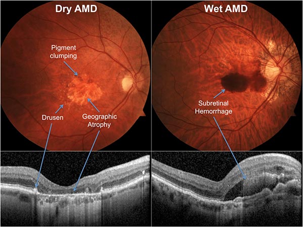
Dry macular degeneration accounts for about 90% of macular degeneration. Drusen builds up in the retina or the retinal pigment epithelium is atrophied. The degree of initial visual impairment varies, and in most cases there is no significant loss of central vision. However, as the photoreceptor cells of the macula gradually atrophy, and as time goes by, the visual acuity gradually decreases and develops into a wet form. Therefore, atrophy and degeneration of the retinal layer and changes in drusen should be regularly followed by an ophthalmologist.
Wet macular degeneration accounts for 90% of blindness. Choroidal neovascularization grows under the retina, and severe visual impairment occurs due to bleeding and exudation caused by the new blood vessel itself. It progresses so quickly that your vision deteriorates rapidly within few weeks. Between several months and years after the disease onset, blindness may occur due to discoid scarring and severe bleeding, and central vision may be impaired due to damage caused by retinal detachment and hemorrhage due to prolonged edema and serous invasion.
For wet macular degeneration treatment, an angiogenesis inhibitor drug is injected directly into the eye every 4 to 6 weeks. In Korea, Avastin and Lucentis injections are representative. Injection therapy is a vascular endothelial growth factor inhibition that blocks vascular leakage and the growth of new blood vessels.
You can expect to maintain and improve vision with injection treatment, but the effect lasts about 4 to 8 weeks, so repeat treatment is necessary. Typically, 3-4 or more infusions may be needed. After the injection treatment, antibiotics should be used to prevent infection with bacteria and the patient should be screened for a certain period of time.
COPYRIGHT ⓒ JULIA LABORATORY. ALL RIGHTS RESERVED. DESIGNED BY MARVEL WORKS
 ENG
ENG  KOR
KOR